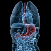
Pleurisy and other pleural disorders affect the pleura, which is a large, thin sheet or membrane of tissue that is wrapped around the outside of the lungs and lines the inside of the chest cavity.
The pleura that line the chest is actually a very thin space that normally holds approximately 4 teaspoons of fluid that is used to keep the two layers of the pleura moving smoothly against one another as a person breathes. If this tissue becomes red or inflamed, that's called pleurisy. The sharp pain that happens with breathing occurs when the two layers of the pleura start rubbing directly against one another.
Pleural Effusion
What is it? & Symptoms - Pleural effusion occurs when there is an abnormal build up of fluid in the pleural space. If the pleural tissues are already inflamed then the fluid will help alleviate the pain associated with the tissues rubbing against each other. But too much fluid can force the layer closest to the lungs against the lungs. Continual build up of fluid and pressure against the lung can cause it to collapse, resulting in difficulty breathing.
Causes - Infections, injuries, heart or liver failure, blood clots in the blood vessels of the lungs (pulmonary emboli), cancer, pneumonia, and medications can all cause the build up of fluid.
Diagnosis - Diagnostic methods usually include chest X-rays, laboratory testing of the fluid (thoracentesis), and a CT scan. The X-rays show the particular areas that are affected by the inflammation. The laboratory testing is needed to determine what kind of fluid has built up and the presence of any bacteria. If there is bacteria build up, the laboratory tests will show what kind of bacteria so the proper antibiotics may be administered. This fluid will also be examined for the amount and types of cells, and for the presence of cancer cells.
Other diagnostic tools include the use of a thoracoscope, which is a tube that when inserted into the chest cavity allows the doctor to visually examine the pleural space and obtain tissue samples. Thoracoscopy can also detect cancer and tuberculosis.
A needle biopsy may be indicated if the equipment for a thoracoscopy is not available. This involves inserting a needle through the chest wall to withdraw a sample of the fluid that has built up. Occasionally, doctors will need to directly view the airways through a viewing tube in a procedure known as a "bronchoscopy" to help them determine the cause of the fluid build up.
TreatmentTreatment of larger infections usually consists of inserting a tube into the chest draining the fluid off, which will relieve the shortness of breath. This is usually done through thoracentesis. Up to 1.5 liters (1.5 quarts) of fluid at a time can be drained using this procedure.
Smaller cases of pleural effusion may not be treated at all, but the doctor will investigate what caused the effusion in the first place, and that will likely be the target of treatment.
Many people do not experience any symptoms at all, but the most common symptom is shortness of breath and chest pain when a person breathes deeply or coughs.
Pneumothorax
What is it? - A pneumothorax happens when there is air - not fluid - between the two layers of pleura. This can cause partial or complete collapse of the lung, leading to extreme shortness of breath and chest pain.
When this happens in a patient that doesn't have any apparent cause or pre-existing lung disorder, it is called a primary spontaneous pneumothorax. It is most common in tall men below the age of 40 who smoke. Although most people fully recover, the condition recurs in up to 50% of people.
Secondary spontaneous pneumothorax occurs in those people with pre-existing lung disorders such as emphysema, cystic fibrosis, asthma, lung abscess, tuberculosis, and Pneumocystis pneumonia.
Causes - A pneumothorax may also result from an injury or medical procedure that introduces air into the pleural space (eg: thoracentesis, bronchoscopy, or thoracoscopy). Ventilators can cause pressure damage to the lungs also resulting in pneumothorax. Those who work or live in a pressurized environment (divers and pilots) may be at increased risk for pneumothorax.
Symptoms - The presence and severity of symptoms varies from patient to patient and depends on the extent of the lung collapse. In addition to shortness of breath, sharp chest pain, and onset of dry, hacking cough, pain may also be felt in the shoulder, neck, or abdomen. A rapidly developing pneumothorax will produce more severe symptoms than a slowly developing case. In most cases, the body will adjust to working without the collapsed lung, and as the fluid is reabsorbed by the body, the lung will slowly reinflate.
Diagnosis - Diagnosis includes a physical examination through a stethoscope and tapping (percussing) of the chest wall. In healthy individuals, the doctor will be able to hear the breathing sounds and a hollow, drumlike sound when tapping on the chest wall. A chest X-ray will be taken to follow up on any abnormal sounds.
Treatment - Minor cases will not require treatment and often do not cause significant breathing problems. In these cases, the air is absorbed within a few days. If breathing is impaired as in a larger case, the air is removed with a large syringe attached to a catheter. If "catheter aspiration" is unsuccessful then doctors will use a chest tube to drain the air. This method is often used for other forms of pneumothorax as well.
Those that experience recurring pneumothorax can have surgery to repair the areas where air is leaking in. Leaving the situation untreated can lead to more infections and increased chronic breathing issues.
Tension Pneumothorax involves the tissues in the area where the air is coming in acting like a pressure valve and not allowing air out. This will completely collapse the lung, and push the heart, and other structures to the other side of the chest. If the pressure is not relieved immediately, death can happen in minutes. In this case, a doctor will use a needle to relieve the pressure and insert a chest tube to continuously drain the air.
Sources: www.nhlbi.nih.gov (U.S. Department of Health & Human Services and National Institutes of Health); www.merck.com


Add a Comment2 Comments
This was an excellent, easy to understand article!
March 13, 2011 - 4:43pmThis Comment
Thank you.
That's what I was aiming for. Some of these conditions are not easy to explain at all!
March 14, 2011 - 6:06amThis Comment