Your breast cancer pathology report may be broken down into the following sections: demographics, gross description/specimen, clinical history/diagnosis, gross description, special tests/markers, microscopic description, prognostic report, summary and diagnosis. Know in advance that different labs and hospitals may use different words to describe the same thing.
Demographics. First, check the top of the report for your name, the date you had your operation and the type of operation you had. Make sure this information is accurate. This section may also include your doctor(s) name(s), medical record number and other identifying information.
Gross Description/Specimen. This section describes where the tissue samples came from. Tissue samples could be taken from the breast, from the lymph nodes under your arm (axilla), or both. In this section, you will find the description of the specimen or tissue sample based on what the naked eye can see. Included here will be the size and weight of the specimen and other visual observations made by the pathologist. Information about how the sample was handled, how it was sectioned and what materials were used in preparation for the microscopic examination is also included.
Clinical History. This is a short description of you and how the breast abnormality was found. It also describes the kind of surgery that was done.
Clinical Diagnosis. This is the diagnosis the doctors were expecting before your breast tissue sample was tested.
Gross Description. This section describes the tissue sample or samples. It talks about the size, weight, and color of each sample.
Special Tests or Markers. This section reports the results of tests for proteins, genes and how fast the cells are growing.
Microscopic Description. This section describes the way the cancer cells look under the microscope. This part of the report points out the features of the tumor or tissue sample. These features will lead to the specific diagnosis.
Prognostic Report. Results of the specific tests performed on the specimen are stated in this section.
Summary and Final Diagnosis. This section is the short description of all the important findings in each tissue sample. Also, this section should contain all of the important data from various tests performed on the specimen leading to a diagnosis. The most helpful part of this section is a list of characteristics and the findings related to the specific specimen.
Another section you may find in the report is staging. Staging is the assessment of how far a patient’s breast cancer has progressed and determines treatment decisions and prognosis. Knowing the patient’s stage helps the doctor decide what kind of treatment would be best for the patient. Staging is done using the TNM system. T is Tumor size, N is lymph Node status (cancer cells found in the lymph nodes is called positive status) and M is Metastasis (the tumor has spread to other parts of the body). In general, the lower the stage the better the prognosis.
• stage 0
• stage I (1)
• stage IIA (2A)
• stage IIB (2B)
• stage IIIA (3A)
• stage IIIB (3B)
• stage IIIC (3C)
• stage IV (4)
Staging is based on the size of the tumor, whether lymph nodes are involved, and whether the cancer has spread beyond the breast. Your doctors use all parts of the pathology report as well as the breast cancer stage to shape your treatment plan.
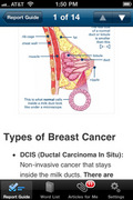
Finally, Breastcancer.org has developed a free breast cancer diagnosis guide for the iPhone, iTouch or iPad. The application is FREE and available at http://itunes.apple.com/us/app/breast-cancer-diagnosis-guide/id389683262?mt=8#.
This app walks you through your breast cancer pathology report and other tests and information that you and your doctor will use to help decide which treatments are right for you.
Add your diagnosis information for easy reference and receive relevant articles featuring the latest research and news about breast cancer. The app includes illustrations and definitions.
Sources:
Breastcancer.org booklet: Your Guide to the Breast Cancer Pathology Report
http://www.networkofstrength.org


Add a CommentComments
There are no comments yet. Be the first one and get the conversation started!